


Spinal osteochondrosis is a chronic disease in which degenerative changes in the vertebral and intervertebral disc located between them occur.Depending on the place of damage to the spine, they distinguish: osteochondrosis of the cervical area, osteochondrosis of the thoracic region and the lumbar area osteochondrosis.To diagnose spinal osteochondrosis, it is necessary to perform radiography, and in the case of its complications (for example, the intervertebral disc hernia) - the spinal MRI.In the treatment of spinal osteochondrosis, along with the drug method, it is widely used, reflexology, massage, manual therapy, physiotherapy and physiotherapy training.
Etiology and pathogenesis
For one degree or another, spinal osteochondrosis develops in all ages and is one of the processes of body aging.Previously, atopic changes occurred in the intervertebral disc, however, injuries, diseases and various spinal benefits contributed to the occurrence of osteochondrosis earlier.The most common osteochondrosis in the cervical region and lumbar spinal osteochondrosis.
About 10 osteochondrosis theories have been developed: vascular, hormone, mechanical, descendants, allergies and more.But not one of them gives a complete explanation of the changes that occur in the spine, but they complement each other.
It is believed that the main point in the occurrence of osteochondrosis is the constant advantage of the vertebral motor segment consisting of two adjacent vertebrae.Such advantages can occur due to motor stereotypes - posture, how individuals sit and walk.Poster disruption, sitting in the wrong pose, walking with an uneven spinal column causes an additional burden on the disc, ligaments and spinal muscles.This process can be worsening due to the characteristics of the spinal structure and the lack of tissue trofism -the genetic factors.Often, evil in the structure is present in the cervical region and leads to vascular disorders and early appearance of signs of cervical spinal osteochondrosis.
The occurrence of osteochondrosis of the lumbar area is more often associated with its burden during tendency and severe elevator.Healthy intervertebral discs can withstand significant load due to the pulpoose nucleus hydrophilic located in the middle.The nucleus contains a large amount of water, and the liquid, as you know, is slightly compressed.Healthy intervertebral disc fractions can occur with compression of more than 500 kg, while the disc changes due to osteochondrosis are ripped by compression of 200 kg.The load of 200 kg suffered a lumbar lumbar of a person weighing 70 kg, when it holds a cargo 15 pounds in a body tilt by 200. The large pressure is due to the low size of the pulp nucleus.With an increase in the tendency to 700, the load on the intervertebral disc will be 489 kg.Therefore, often the first clinical manifestation of lumbar spinal osteochondrosis occurs during or after weight lifting, doing homework, grass in the garden, etc.
Destruction of connective tissue of the discs, ligaments and capsules of the facet joints causing the reaction of the immune system and the development of aseptic inflammation with the swelling of the facet joints and surrounding tissues.Due to the body's displacement of the vertebral body, the facet joint capsule is stretched, and the altered intervertebral disc is not very strong by the bodies of the neighboring vertebrae.The instability of the spinal segment is formed.Due to instability, infringement of the spinal cord with the development of radicular syndrome is possible.With cervical spinal osteochondrosis, this often occurs during rotation of the head, with lumbar area osteochondrosis - during the body's tilt.It is possible to form the function of the vertebral motor segment.It is caused by a reduction in vertebral muscle compensation.
The intervertebral disc hernia is formed when the disc returns, the posterior longitudinal ligaments rupture and the disk enlargement of the spinal cord occur.If at the same time the nucleus of the disc is cooked on the cerebrospinal tract, then such a hernia is called explosion.The severity and duration of pain with such hernia are much larger than with the unexpected.Disc hernia can cause radical syndrome or spinal cord compression.
With osteochondrosis, bone tissue growth occurs with the formation of osteophytes - growing bone in the body and the vertebral process.Osteophytes can also cause spinal cord compression or cause the development of radicular syndrome.
Symptoms of spinal osteochondrosis
The main symptom of spinal osteochondrosis is pain.Pain can be acute with high intensity, it is increasing with a slight movement in the affected segment and thus makes the patient take forced.Therefore, with cervical spinal osteochondrosis, the patient holds his head in the most painless and unable to change it, with osteochondrosis from the thoracic region, pain increases even with deep breathing, and with osteochondrosis in the lumbar area, difficult to sit, up and walk.Such pain syndrome is a characteristic of compressing the spinal cord.
In about 80% of cases, there is a tedious pain of persistent nature and moderate intensity.In such cases, after the examination, the doctor must distinguish the manifestation of spinal osteochondrosis from the back myositis of the muscles.Stupid pain in osteochondrosis is due to excessive muscle tension, holding the affected vertebral motor segment, changes in inflammation or significant intervertebral disc.In patients with such pain, the position must be absent, but movement restrictions and physical activity are lowered.Patients with cervical spinal osteochondrosis avoid sharp turns and tilt with their heads, with osteochondrosis in the lumbar region - slowly sitting and waking, avoid leaning.
Complications of spinal osteochondrosis
Complications of osteochondrosis are associated with intervertebral disc hernia.These include the compression of the spinal cord, characterized by numbness, weakness of certain muscle groups from the feet (depending on the level of compression), leading to the appearance of paresis, muscle atrophy, changes in tendon reflex, urine and water removal.Intervertebral hernia can cause arterial compression that consumes spinal cord with the formation of ischemia (spinal cord infarction) with nerve cell death.This is reflected in the emergence of neurological deficits (affected movements, sensitivity, trophic disorders) that are equal to the level and prevalence of ischemia.
Diagnosis of spinal osteochondrosis
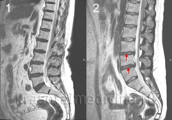
The diagnosis of spinal osteochondrosis is performed by a neurologist or vertebrologist.In the early stages, spinal radiography was performed in 2 projections.If necessary, they can shoot separate spinal segments and shoot in additional projections.For the diagnosis of intervertebral hernia, evaluate the spinal cord and detect complications of osteochondrosis, magnetic tomography and resonance (spinal MRI) are used.An important role is played by MRI in the diagnosis of differential osteochondrosis and other spinal diseases: spondylitis tuberculosis, osteomyelitis, tumor, spondel ankylosing, rheumatism, infectious wounds.Sometimes in the case of complicated cervical spine osteochondrosis, the exception of syringomyelia is required.In some cases, if MRI is impossible, myelography is indicated.
The targeted study of the affected intervertebral disc is likely to use diskography.Electrophysiological studies are used to determine the level and localization of damage to the nerve path, to monitor their recovery process during therapy.
Treatment of spinal cord osteochondrosis
During the acute period, peace is shown in the affected vertebral motor segment.For this purpose, with cervical spinal osteochondrosis, fixation is used using chantz collar, with osteochondrosis in the lumbar area - bed break.Setting is also required for cervical area osteochondrosis with the instability of the vertebral segment.
In osteochondrosis drug therapy, anti -non -ssteroid (NSAIDs) anti -inflammatory drugs (NSAIDs) are used: diclofenac, nimesulide, lornoxicam, meloxicam.With intense pain syndrome, analgesics are shown, for example, flapportin analgesic center action.To relieve muscle voltage, muscle relaxants are used - tolperisone, thizanidine.In some cases, it is advisable to prescribe anticonvulsant - carbamazepine, gabapentin;Antidepressants, including priority given to serotonin arrest inhibitors (cerseralin, paroxetine).
In the event of radicular syndrome, the patient's treatment is indicated.It may be local introduction of glucocorticoids, treatment for edema, the use of attractiveness.In the treatment of osteochondrosis, physiotherapy, reflexology, massage, physiotherapy training is widely used.The use of manual therapy requires a clear compliance with its implementation techniques and caution in the treatment of cervical spinal osteochondrosis.
Spinal surgery is indicated primarily with significant spinal cord compression.It consists of eliminating intervertebral disc hernia and spinal cord compression.It is possible to perform microdisectomy, laser disk reconstruction, disk replacement of the implant, stabilization of the spinal segment.